Pathology is becoming a computational discipline. The models get the headlines - but the value is decided by the data foundation beneath them.
Key takeaways
- AI is turning pathology from a microscope discipline into a computational one - yet the breakthrough that gets attention, the models, sits on a foundation most laboratories underestimate.
- Digital pathology is becoming enterprise infrastructure, not a lab gadget: whole-slide imaging at scale demands serious storage, standards-based integration, security and governance.
- Today's approved tools augment pathologists rather than replace them - every clinical decision still needs human sign-off, and every model needs assurance and monitoring.
- The organisations that benefit most are those that get the data foundation - image data, standards, identity, quality and governance - right first.
The microscope becomes a computer
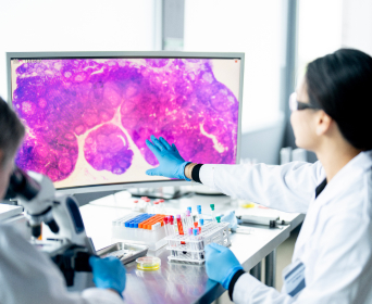
For roughly 150 years, pathology has been a visual discipline: a trained physician examines stained tissue under a microscope and renders a diagnosis from pattern recognition. That is changing fast. The glass slide is becoming a file. Whole-slide imaging digitises tissue at gigapixel resolution, and AI is learning to read those pixels - flagging regions of interest, quantifying biomarkers and supporting prognosis.
In 2026 the pace is striking: pathology-specific foundation models, a growing list of regulatory clearances, and AI copilots that assist diagnosis and reporting. The temptation is to read all this as an algorithm story. It is not. The algorithm is the visible tip of a much larger iceberg - and the value depends on the data and infrastructure beneath it. That is where the real work, and VE3's focus, sits.
What is computational pathology?
Computational pathology is the application of AI to digitised tissue images to detect, quantify and predict - supporting the pathologist's diagnosis. It builds on digital pathology, which is the digitisation of glass slides into whole-slide images (WSIs) so they can be viewed, shared and stored electronically. One is the medium; the other is the intelligence layered on top of it.
The distinction matters. Many laboratories have begun digitising slides without yet building the data foundation that computational pathology requires - and it is that gap, not the availability of algorithms, that most often stalls progress.
Why is AI in pathology accelerating now?
Several forces are converging at once.
- Whole-slide imaging has matured. Scanners, viewers and storage are now capable of enterprise-scale digitisation, and remote, collaborative diagnostics have become a practical necessity rather than an experiment.
- Foundation models have changed the economics. Pathology-specific foundation models from academic and commercial labs, pre-trained on very large image collections, can be adapted to new tasks with a fraction of the labelled data once required - lowering the barrier to new applications.
- Multimodal AI and copilots are arriving. Systems that combine imaging with other data, and LLM-based assistants that help with reporting and workflow, are moving from research into support tools.
- Regulators are moving with the field. Approved AI tools now span areas such as prostate cancer detection, cervical cytology screening and biomarker quantification (for example HER2 and PD-L1), with newer tools receiving expedited designations - a clear shift from experimental to clinically relevant.
- Workforce and demand pressure. Rising caseloads and pathologist shortages make efficiency, case prioritisation and remote collaboration compelling, while reimbursement pathways for diagnostic AI are beginning to emerge - so adoption is no longer waiting on perfect economics.
One-point cuts through the hype: every currently approved tool augments the pathologist. None replaces them, and all require final human sign-off. The direction of travel is decision support, not autonomy.
The model is the easy part
Here is the truth that vendor demonstrations tend to gloss over: computational analysis is only as good as the tissue - and the data - it reads. A single whole-slide image is enormous; one case can span multiple slides; and a busy laboratory produces these at volume, every day.
Behind every model, therefore, sits an infrastructure problem. Gigapixel images must be captured, stored and moved at scale, with archival retention and real cost implications. They must be standardised so that scanners, viewers, the laboratory information system (LIS) and analytics tools all interpret them consistently - which is what DICOM (image encoding), HL7 (metadata) and the IHE Digital Pathology acquisition profile exist to enable. Every image must be linked reliably to the right patient and case. And access to some of the most sensitive data in medicine must be governed throughout.
This is why digital pathology in 2026 is increasingly rolled out less like a standalone lab upgrade and more like core enterprise infrastructure. That same foundation increasingly underpins how the models themselves are built - through large, well-curated image repositories and privacy-preserving, federated approaches that let institutions collaborate without moving sensitive data. Get the foundation wrong, and even the most capable model will produce unreliable results - confidently. Get it right, and the same model becomes dependable, repeatable and safe to build on.
From digital slides to decision support: what good looks like
The laboratories turning pathology AI into trustworthy decision support treat the foundation as seriously as the algorithm. Five capabilities do the work.
- A scalable image data platform - storage and compute sized for gigapixel WSIs and long-term retention, instrumented for cost control from the outset.
- Standards-based integration - DICOM, HL7 and IHE profiles so scanners, the LIS, viewers and analytics interoperate rather than lock the lab into a single vendor.
- Identity and data quality - reliable linkage of slides, cases and patients through entity resolution, so every result attaches to the right person.
- Multimodal linkage - connecting pathology images with genomic and clinical data to support precision oncology, where the diagnosis increasingly depends on more than the slide alone.
- Governed access and assurance - security, audit and consent controls over sensitive tissue data, feeding only validated models.
Built this way, pathology AI stops being a series of isolated pilots and becomes a capability the whole laboratory can rely on and extend.
Augment, not replace: the assurance that makes it deployable
Governance is not a footnote here - it decides whether pathology AI reaches the bench at all. Because these tools inform clinical decisions, they are regulated as software as a medical device. In the UK that means MHRA classification, clinical-safety cases (DCB0129 and DCB0160), and DTAC and NICE evidence expectations; internationally, the FDA and EU routes apply.
Approval is only the start. Deployed models need real-world performance monitoring, because variation in tissue preparation, staining and scanners causes drift that can quietly erode accuracy over time. A clinical-AI assurance approach - covering validation, human oversight and ongoing monitoring - is what allows a laboratory to adopt AI with confidence rather than caution. Handled well, assurance is not a brake on innovation; it is the enabler that lets a lab say yes.
A pragmatic path forward
There is no need to digitise and automate everything at once. A more effective route is incremental: digitise the highest-value workflows first; build the image data platform and standards-based integration beneath them; prove a single AI use case end to end under proper assurance; then scale, and connect pathology data to molecular and clinical records. Each step should deliver usable value, not just infrastructure.
Foundations first
The move from digital slides to decision support is one of the most significant shifts in modern diagnostics. But it will be won on foundations, not on algorithms alone. The laboratories that pull ahead will be those that can capture, standardise, connect, govern and trust their image data - and then let AI do its work on top of a foundation built for it.
VE3 helps diagnostics and pathology teams build exactly that: scalable, standards-based, governed image-data foundations, with the data quality and clinical-AI assurance that make AI-assisted diagnosis dependable. If you are digitising pathology and want the foundation to match the ambition, talk to our healthcare data team.